

This image shows what finding? What might it indicate?
GI Case 1
Autor: Marc Harth - Institution: Uniklinik Frankfurt Institut für Diagnostische und Interventionelle Radiologie
Abstract: 46 year old female with 2 days of right sided abdominal pain.
Examination
46 year old female with 2 days of right sided abdominal pain.
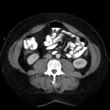
This is an image from a contrast enhanced abdominal CT.
 "Zoom (34KB)")
These are images from a contrast enhanced abdominal CT.
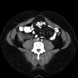
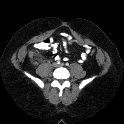
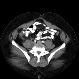
Findings
Fluid in the right paracolic gutter, and a thickened, inflamed appendix with thickening of the adjacent cecal wall.
Differential: Acute appendicitis is by far the most likely diagnosis.
No appendicolith is seen in this case.
Diskussion
The appendix is normally less than 7 mm in diameter, or if fluid filled, the wall can be up to 3mm thick. This appendix is distended, and the adjacent stranding and fluid indicates the inflammatory process which is causing peritoneal irritation and pain. Here is a series of images showing a normal appendix using similar technique. Note the thin walls of the normal appendix.
Neoplastic processes can obstruct the appendiceal orifice, causing buildup of fluid and secretions in the obstructed portion. Distention will eventually slow or stop venous outflow, and cause venous stasis and inflammation. Appendiceal mucocele could also act in a similar manner.
Quiz
Findings of Appendicitis on CT can include all except:
cecal wall thickening
gastric wall thickening
periappendiceal fat stranding
